Back and scalp lesions in the newborn
Please note: images that have a white symbol at the top right, such as the Sacral Dimples image below, indicates an image gallery that has multiple images - click on the image to open the gallery.
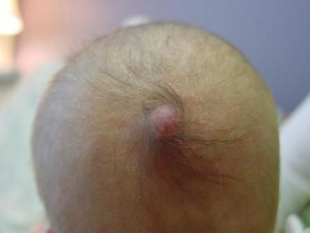
Midline scalp lesions

Lesions over the midline scalp require particular attention. They may represent lesions which communicate with underlying nervous system structures (encephalocoeles or meningocoeles) or other abnormalities of nervous system tissue (heterotopic meningeal or neural tissue).
Typically, there is a collar of hair surrounding the lesion. The hair is usually darker, more coarse, and longer than the other scalp hair. There may also be an associated capillary malformation adjacent to or over the lesion. Heterotopic neural tissue may also be seen away from the scalp, such as on the face.
It is important to undertake imaging to determine if the lesion communicates with underlying structures, particularly before considering surgical removal.
Sacral dimples
Sacral dimples are relatively common, occurring in 2-4% of newborn infants. They may be associated with a tuft of hair. Almost always, if the dimple is within the gluteal crease, there is no underlying spinal abnormality and no investigation is necessary.
Dimples that may require further investigation are those that are large (>5mm), deep (may represent dermal sinuses and may communicate with the underlying spinal canal), those above the gluteal crease, or those associated with other cutaneous markers of spinal dysraphism (as below).
The deeper lesions may be prone to infections secondary to trapped debris and hair etc. The parents should be advised to maintain local hygiene and to teach the child the same at a later stage.
In the neonatal period, an ultrasound may give pictures which demonstrate any communication with the cutaneous abnormality. The last image to the right shows a normal spinal cord as imaged above the lesion; no communication is normal, and the spinal cord is seen tapering.
Midline skin lesions of the lower back
The first series of photographs depict a large hairy patch overlying the midlumbar region. Plain radiographs revealed thoracic vertebral segmentation defects. In the lumbar region, increased interpediculate distance, flattening of the vertebral bodies and narrowing of the intervertebral spaces, as well as the presence of a calcified "spur" on the lateral view were consistent with diastematomyelia. Ultrasound showed a split cord.
This is a typical example of the relationship between superficial skin lesions overlying the midline of the back and underlying spinal anomalies. Any lesion from the lower lumbar region to the upper cervical region that covers the midline should be treated with respect. Consider the possibility of an underlying spinal anomaly until proven otherwise and obtain the necessary investigations: plain films and ultrasound.
In contrast, sacral dimples, pits, or sinuses present within the intergluteal cleft are common benign lesions thought to occur in between 2% and 4% of newborn babies. They do not require investigations provided that there are no associated concomitant lesions higher up and no evidence of orthopaedic or neurologic anomalies of the lower limbs. Parents should be advised on maintaining local hygiene as the deeper lesions may eventually harbour debris and hair with possibility of secondary infection.
This is another example of a midline lesion above the sacral cleft. This had a soft lipomatous feel on palpation. On close inspection a sinus opening was noted hidden underneath the central lipomatous mass. The infant had no lower limbs anomalies, exhibited normal movements and normally brisk and equal deep tendon reflexes. Ultrasound examination revealed a lipomatous mass with attachment to the dura and associated tethering of the spinal cord.
