Antenatal Doppler Studies in High Risk Pregnancies
Background
Doppler ultrasound detects frequency shifts from moving targets (red blood cells). The flow velocity waveform (FVW) which is produced is unique to the vessel being studied.
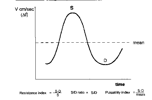
Flow velocity waveforms are analysed mathematically by creating a ratio of the systolic to diastolic frequencies. The most common index in use at NWH is the resistance index (see figure 1).

Figure 1: Maximum frequency envelope of Doppler flow velocity waveform showing peak systolic frequency shift (S) and end-diastolic frequency shift (D).
Umbilical artery
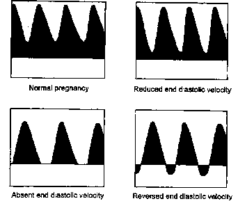
In normal pregnancy, there is a progressive increase in end-diastolic velocity due to growth and dilatation of the umbilical circulation. The resistance index therefore falls. A resistance index > 0.72 is outside the normal limits from 26 weeks gestation onwards. In some pregnancies with fetal growth restriction and/or preeclampsia, there is a reduction in the diastolic velocity and in severe cases as in c) and d) below, there is absent or reversed end diastolic velocity.

Figure 2: Umbilical Doppler waveforms in healthy pregnancy and in pregnancies with fetal growth restriction.
These abnormal waveforms are associated with histological evidence of reduced numbers of small placental blood vessels and therefore reflect "placental insufficiency". In pregnancies with reduced, absent or reversed end-diastolic velocity, there is an increased risk of stillbirth, asphyxia, chromosomal and congenital abnormality. Abnormal umbilical Doppler waveforms can be present for weeks before there is evidence of fetal compromise and therefore are a marker of a high risk situation and should not normally be used in isolation as an indication for delivery. However, most obstetricians would consider delivering a fetus with absent end-diastolic velocity from about thirty two weeks gestation following administration of corticosteroids. In many instances, fetuses with absent end-diastolic velocity would present much earlier in pregnancy and may require extremely preterm delivery.
Umbilical artery Doppler studies have been well evaluated in randomised controlled trials and use of umbilical artery Doppler studies in small for gestation age pregnancies and in pregnancies with the preeclampsia is associated with a 30% reduction in perinatal mortality (Cochrane Library 2000). The likely reason for the reduced perinatal mortality is that the abnormal Doppler waveform highlights the at risk fetus who is then subject to more frequent surveillance ultimately resulting in timely preterm delivery.
About two thirds of small gestational age fetuses have normal umbilical artery Doppler studies and are unlikely to have histologic evidence of abnormal placental vascular development . These fetuses have a very low risk of antenatal fetal compromise, have a low perinatal mortality, and do not normally require preterm delivery.
Uterine Artery Doppler Studies
Uterine artery Doppler studies reflect the resistance in the utero-placental circulation and have not been proven to reduce perinatal morbidity or mortality. In conditions such as preeclampsia and fetal growth restriction abnormal uterine artery Doppler studies indicate inadequate placentation and have been correlated with histological evidence of defective replacement of spiral arteries by maternal trophoblast.

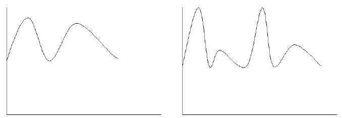
| Normal Uterine Artery Doppler | Abnormal Uterine Artery Doppler |
Abnormal uterine artery Doppler studies in the context of a growth restricted pregnancy suggest a maternal cause for the growth restriction where as normal uterine artery Doppler studies suggest that a fetal cause of growth restriction needs to be considered.
