Umbilical catheters in the neonate

Please note: images that have a white symbol at the top right, such as the UAC in the high position image below, indicates an image gallery that has multiple images - click on the image to open the gallery.
UAC in the wrong vessel and UVC in the wrong vessel
Be careful to identify the vessels correctly.
The image to the right shows the double lumen venous catheter placed in the aorta, and the supposedly arterial catheter inserted into the UVC and - being sited high - in the jugular vein.
This tends to make the gas you take off the "arterial" catheter a bit worse than you expected ....
UAC in the high position / too high
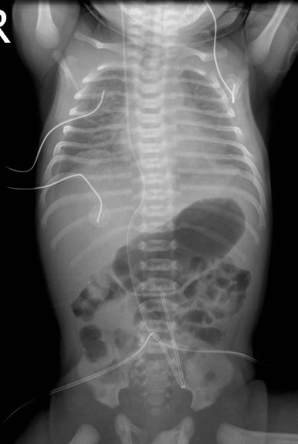
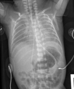
In the first two images the UAC is at T6, which is satisfactory. The UVC is at T7. Ideally, both should be above the diaphragm. The UAC should be between T6 and T9¹. The UVC should be in the IVC as it enters the right atrium.
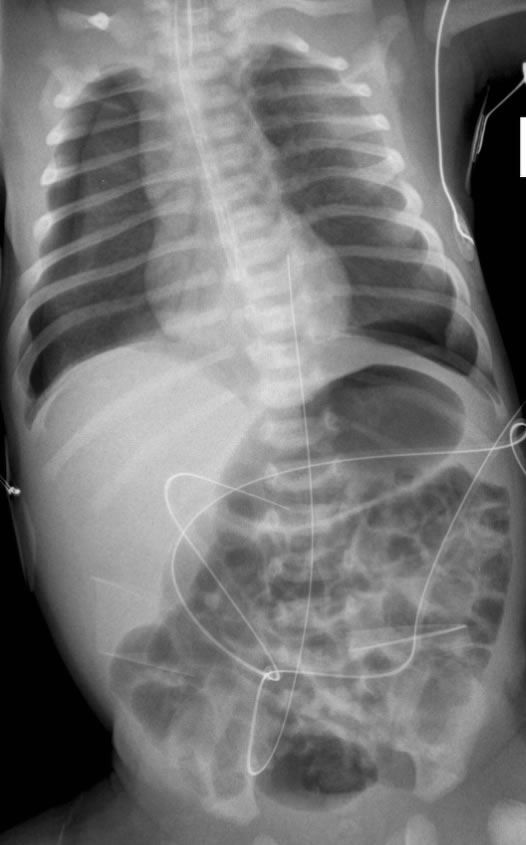
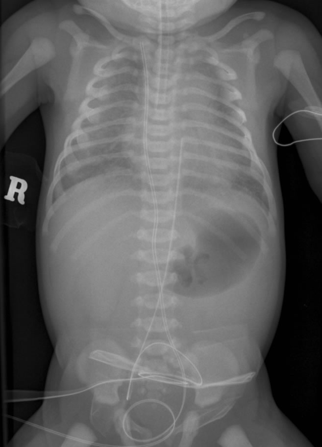
The third and fourth images show the UAC clearly in too high, sitting just below the left subclavian artery.

UAC in the left subclavian artery
This UAC has been inserted far too deeply in a small baby and has ended up in the left subclavian artery.
Not surprisingly, it was not reading the blood pressure too well, nor sampling well.
The UVC probably just enters the right atrium.
UAC not in far enough
Two UACs are in place, one in the right iliac artery, the other in the lower aorta at the level of the upper border of L2.
Neither position is satisfactory. The left catheter could be in a satisfactory position if withdrawn to L3/4. ¹
UAC in main pulmonary artery (via PDA)
On the AP film, the UAC looks high at T4.
Clinically, the baby looked very pink and was saturating at 100%, but the PaO2 on a blood gas through the UAC showed a PaO2 of only 5kPa.
On the lateral radiograph, the UAC does not follow the expected course of the aorta and is too anterior. The UAC follows the PDA and the tip is in the main pulmonary artery.
The UVC is also in too far, and interestingly is probably in the left atrium, thereby giving a better indication of PaO2 than the UAC.
UAC in the abdomen (somewhere?)
The UVC is in the right atrium, the tip is perhaps a little high, close to the atrial septum.
The UAC is looped upon itself and is probably tracking subcutaneously.
UACs in the gluteal arteries
Two UACs have been inserted. Neither is in an appropriate position. Both enter the umbilical arteries and seem to track posteriorly, possibly into the gluteal arteries. Contrast has been injected into one of the catheters.
The UVC is in a satisfactory position.
UVC too low
The tip of the UVC lies below the diaphragm (too low), though not in the liver.
The UAC is positioned too high tending towards the left subclavian artery.
UVC in liver
The UVC is coiled and has its tip projected over the right upper quadrant, most likely in the liver.
The UAC has its tip at the expected position of the left common iliac artery.
UVC looped in the liver
The UVC on the AP does not appear to be in far enough and is lying in the liver.
On the lateral it is clearly looped anteriorly.
The UAC is also slightly high at T5.
In the images to the right, the UVC is also coiled in the liver, but appears to make a correct turn around and exit via the ductus venosus. The coil is probably within the capacious space where the umbilical vein and left portal vein join.
Note is made that the UAC is high at T3.

UVC in portal vein
The UVC is kinked and lies within the left portal vein in aberrant position.
The UAC is in satisfactory position at the level of T8.

UVC in too far (into pulmonary vein)
The tip of UVC has passed across the foramen ovale and likely into the left pulmonary vein.
The UAC has its tip at T3/4 interspace (too high).

UVC in too far (into jugular vein)
The UVC is too high at the level of the confluence of jugular and subclavian veins.
The UAC is in satisfactory position at the level of T6/7 disc space.
UVC in too far (into left ventricle)
The UVC is in too far, passing through the foramen ovale and into left atrium then left ventricle.
The UAC is at T9.
Reference
Fletcher MA, MacDonald MG, Avery GB (Eds). Atlas of procedures in neonatology. JB Lippincott Co, Phil.
Thanks to Dr Jutta van den Boom for collating some images.
